


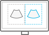
ClearVision enhances the edge contrast and creates sharp 2D images for optimal diagnostic performance.
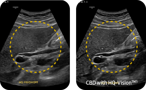
HQ-Vision™ ¹ provides clearer images by mitigating the characteristics of ultrasound images that are slightly blurred than the actual vision.
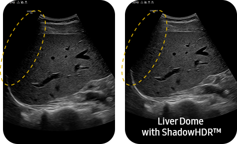
ShadowHDR™ selectively applies high-frequency and low-frequency of ultrasound to identify shadow areas where attenuation occurs.
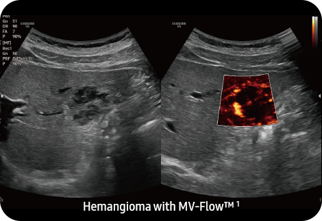
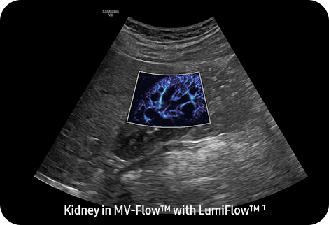
MV-Flow™ ¹ visualizes microcirculatory and slow blood flow to display the intensity of blood flow in color.
LumiFlow™ ¹ is a function that visualizes blood flow in 3 dimensional-like to help understand the structure of blood flow and small vessels intuitively.
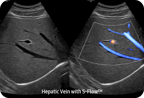
S-Flow™, a directional Power Doppler imaging technology, can help to detect even the peripheral blood vessels. It enables accurate diagnosis when the blood flow examination is especially difficult.

S-Shearwave Imaging™ ¹ allows the non-invasive assessment of stiff tissues in various applications. The color-coded elastogram, quantitative measurements, display options, and user-selectable ROI functions are useful for accurate diagnosis.
.
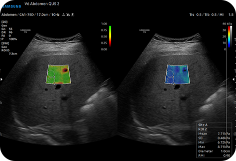
TAI™ ¹ (Tissue Attenuation Imaging) provides quantitative tissue attenuation measurement to assess steatotic liver changes.
TSI™ ¹ (Tissue Scatter distribution Imaging) provides quantitative tissue scatter distribution measurement to assess steatotic liver changes.

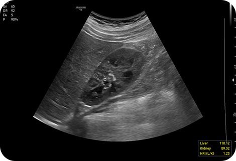
HRI (Hepato Renal Index) is an index to quantify steatosis of a liver by comparing echogenicity between liver parenchyma and renal cortex. EzHRI™ ¹ places 2 ROIs on the liver parenchyma and renal cortex and provides HRI ratio.
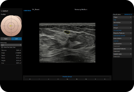
S-Detect™ ¹,³ for Breast analyzes selected lesions in the breast ultrasound study and shows the analysis data, applies BI-RADS ATLAS* to provide standardized reporting; and helps diagnosis with the streamlined workflow.
S-Detect™ ¹,³ for Thyroid analyzes selected lesions in the thyroid ultrasound study and shows the analysis data, provides standardized reporting based on the ATA, BTA,EU-TIRADS, K-TIRADS, and ACR-TIRADS* guidelines; and helps diagnosis with the streamlined workflow.
E-Strain™ ¹,² is designed to enable quick and easy calculation of the strain ratio between two regions of interest for day-to-day practice. Simply by setting the two targets, you can receive accurate, consistent results and make informed decisions in many types of diagnostic procedures.
A diagnostic ultrasound technique for imaging elasticity, ElastoScan+™ ¹ observes the transformation of the tissue strain by the internal or external forces, and converts relative stiffness into a color image.
CEUS+ ¹ is a contrast agent imaging technology. The micro-bubble contrast agent injected into the body through the vein or alike is subjected to perform nonlinear resonance due to stimulation of ultrasound energy.
HeartAssist™ ¹, a feature based on Deep Learning technology, provides automatic classification of ultrasound image into measurement views required for heart diagnosis and provides measurement results.

Strain+ ¹ is a quantitative tool for measuring global and segmental wall motion of the left ventricle (LV). Three standard LV views and a Bull’s Eye are displayed in a quad screen for easy assessment of the LV function.

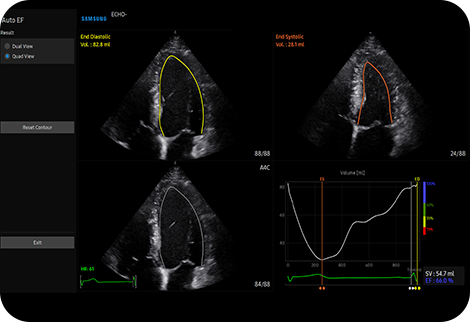
AutoEF ¹ is a feature which conveniently measures and quantifies Ejection Fraction. By selecting the three points of the left ventricle, the volume at the end-systolic and end-diastolic points of the left ventricle is calculated, to assist in quick and efficient assessment of the heart function.
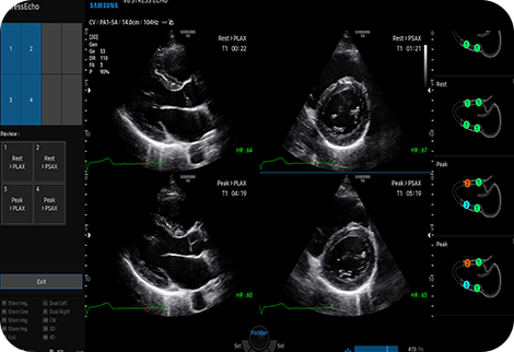
StressEcho ¹ package includes wall motion scoring and reporting. It includes exercise StressEcho, pharmacologic StressEcho, diastolic StressEcho and free programmable StressEcho.
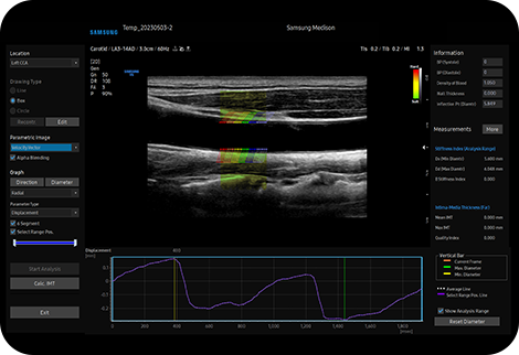
AutoIMT+ ¹ is a screening tool to analyze a patient's potential risk of cardiovascular disease. It allows easy intima-media thickness measurement of both the anterior and posterior wall of the common carotid by the click of a button.

ArterialAnalysis™ ¹ detects functional changes of vessels, providing measurement values such as the stiffness, intima-media thickness and pulse wave velocity of the common carotid artery. Since the functional changes occur before morphological changes, this technology supports the early detection of cardiovascular disease.
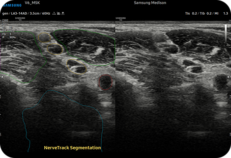
NerveTrack™ ¹, a feature based on Deep Learning technology, detects and provides information of the location of the nerve area in real-time during ultrasound scanning.
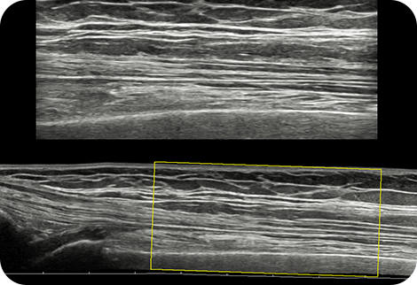
Panoramic+ ¹ imaging displays as an extended field-of-view so users can examine wide areas that do not fit into one image as a single image. Panoramic+ imaging also supports angular scanning from linear transducer data acquisition.
With pinpoint precision, NeedleMate+™ ¹ delineates needle location when performing interventions such as nerve blocks. Improved accuracy and efficiency in procedure are possible with beam steering added to NeedleMate+™ ¹.

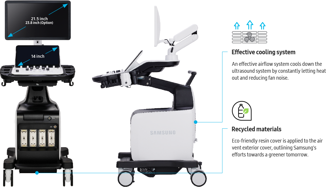
BatteryAssist™ ¹ provides battery power to the system, enabling users to perform scans when AC power is temporarily unavailable. It also allows the system to be moved without having to turn the power off and then back on.

The ultrasound examination can be performed while viewing the images and cines that are expanded at various ratios according to the user preference.

EzExam+™ ¹ enables you to build or use a predefined protocol, and assign protocols for examinations that are regularly performed in the hospital in order to reduce the number of steps that you have to go through.
TouchEdit, a customizable touchscreen, allows the user to move frequently used functions to the first page.
EzCompare™ automatically matches the image settings, annotations, and bodymarkers from the prior study.
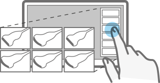
QuickPreset allows the user to select the most common transducer and preset combinations in one click.
The buttons around the trackball can be customized for easy selection of commonly used functions.
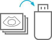
User can directly export image/cine with a USB device.
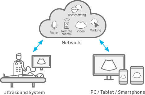
SonoSync™ 1, 5 is available in PC and smartphone, etc. as a real-time image share solution that allows communication for care guide and training between doctors and sonographers. In addition, voice chatting, text chatting and real-time marking functions are provided for better communication; and the MultiVue function is included that allows monitoring multiple ultrasound images on a single screen.
--- --- --- --- --- --- --- --- --- --- --- --- --- --- --- --- --- ---
Saaeda: The region’s most trusted and well-known supplier of healthcare solutions & medical equipment

We are keen to train our employees to be ready to serve our valued customers and We also train our customers on the optimal use of our devices so that they can benefit as much as possible from the products features which will let them develop their healthcare business

We take in consideration warehousing & temperature conditions (Cold Chain) when transport our equipment as these items need an extra level of care.
Our customers rely on us for accurate results and diagnosis, so we make sure to deliver what achieve the confidence you need.
--- --- --- --- --- --- --- --- --- --- --- --- --- --- --- --- --- ---