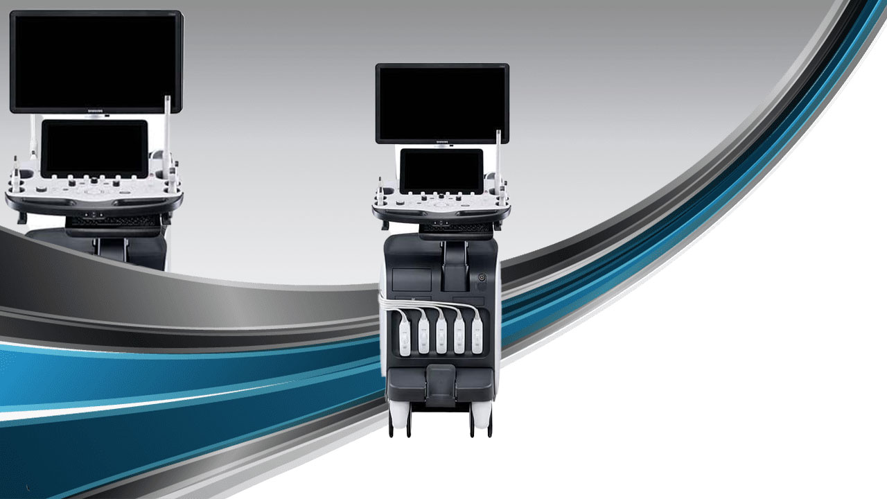

A Revolutionary Change inAdvanced Diagnostics
RS85 Prestige has been revolutionized with novel diagnostic features across each application based on the preeminent imaging performance. The advanced intellectual technologies are to help you confirm with confidence for challenging cases, while the easy-to-use system supports your effort involved in the routine scanning. Especially, Samsung ultrasound's largest
27-inch OLED monitor enhances the diagnostic confidence of healthcare professionals by providing clear and stunning image quality.
Crystal Architecture™, an imaging architecture that combines CrystalBeam™ and CrystalPure™,while based upon S-Vue Transducer™, is to provide crystal clear image.
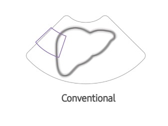


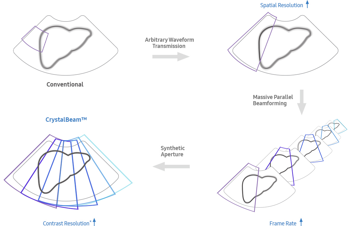
CrystalPure™ imaging engine helps you to make more confident diagnoses with fundamental 2D images and enhanced color performance. It also lessens the incidence of clutter and boosts the level of color signal processing.
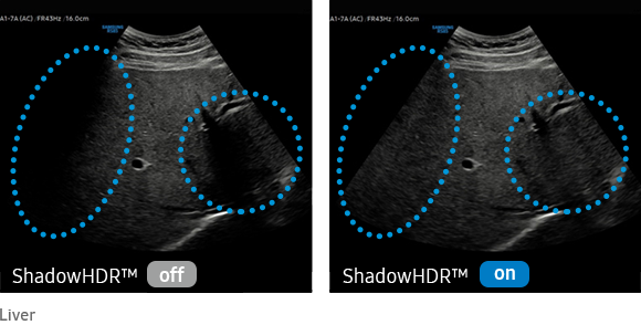
ShadowHDR™ selectively applies high-frequency and low-frequency ultrasound to identify shadow areas where attenuation occurs.
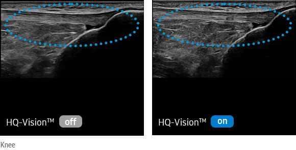
HQ-Vision™ provides clearer images by mitigating the characteristics of ultrasound images that are slightly blurred than the actual vision.
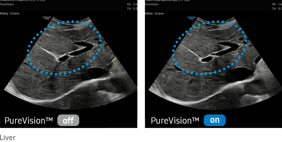
PureVision™ is an image processing function that outputs with a good uniformity and clear image by performing speckle noise suppression and edge enhancement
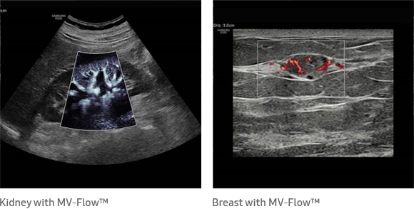
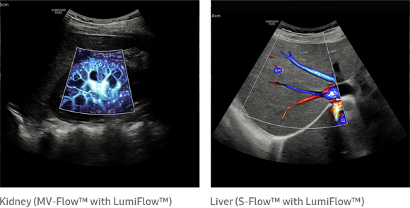
MV-Flow™ ¹ visualizes microcirculatory and slow blood flow to display the intensity of blood flow in color.
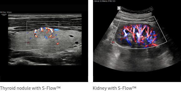
S-Flow™, The function uses directional power doppler technology, enabling you to examine even the peripheral vessels. It displays information on the intensity and direction of blood flow.
LumiFlow™ ¹ is a function that visualizes blood flow in 3 dimensional-like to help understand the structure of blood flow and small vessels intuitively.
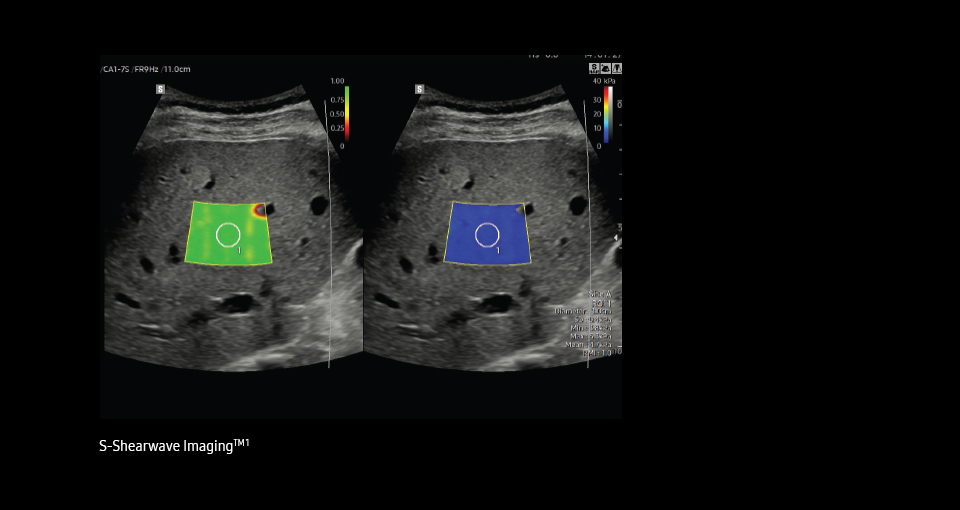
Our features enable healthcare professionals to navigate and quantify ultrasound propagation in realtime,helping them to visualize and make their assessments with accuracy.
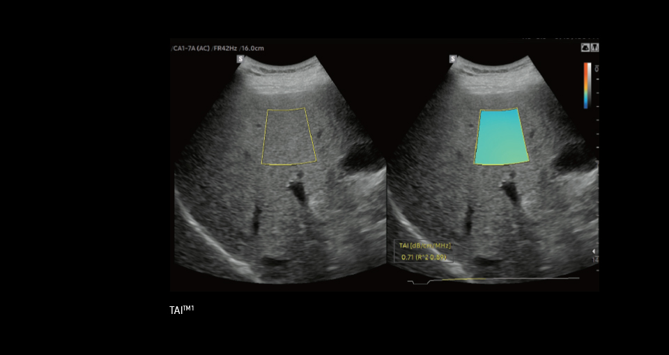
TAI™ ¹provides a quantitative tissue attenuation measurement
to assess steatotic liver changes.
TSI™ ¹ provides a quantitative tissue scatter distribution
measurement to assess steatotic liver changes.
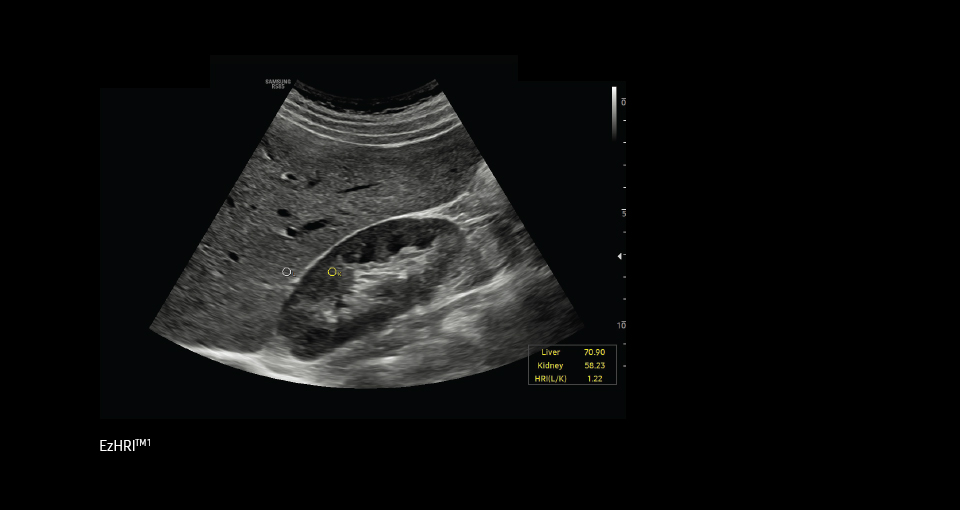
HRI (Hepato Renal Index) is an index to quantify steatosis of a liver
by comparing echogenicity between liver parenchyma with renal
cortex. EzHRI™ ¹ places 2 ROIs on the liver parenchyma and renal
cortex and provides HRI ratio.
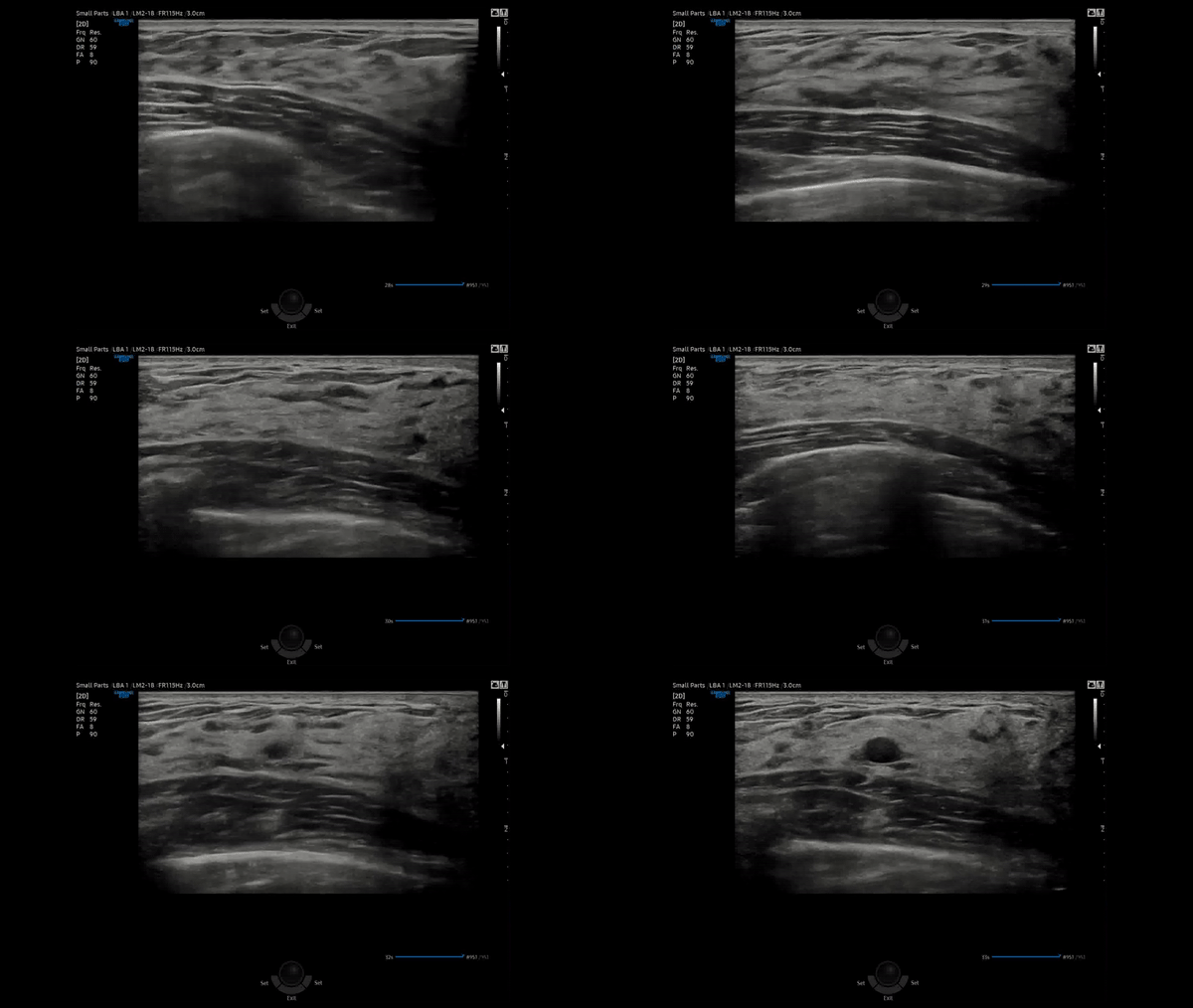
Live BreastAssist™ ¹ , a feature based on Deep Learning technology, detects interested areas in real-time during breast scanning and displays the location of lesions to assist healthcare professionals in diagnosis.
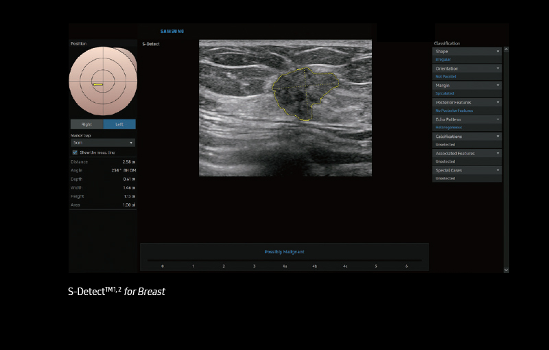
S-Detect™ ¹, ² for Breast analyzes selected lesions in the breast ultrasound study and shows the analysis data, applies BI-RADS ATLAS* to provide standardized reporting; and helps diagnosis with the streamlined workflow.
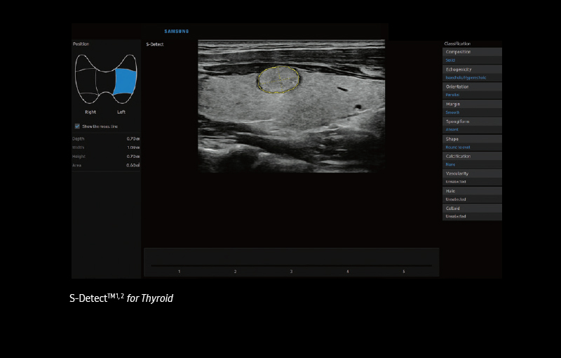
S-Detect™ ¹, ² for Thyroid analyzes selected lesions in the thyroid ultrasound study and shows the analysis data, provides standardized reporting based on the ATA, BTA, EU-TIRADS, K-TIRADS, and ACR TI-RADS guidelines; and helps diagnosis with the streamlined workflow.
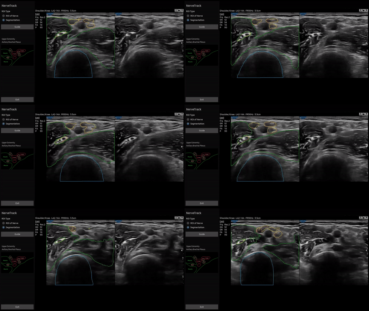
NerveTrack™ ¹ , a feature based on Deep Learning technology,
detects and provides information of the location of the nerve area
in real-time during ultrasound scanning.
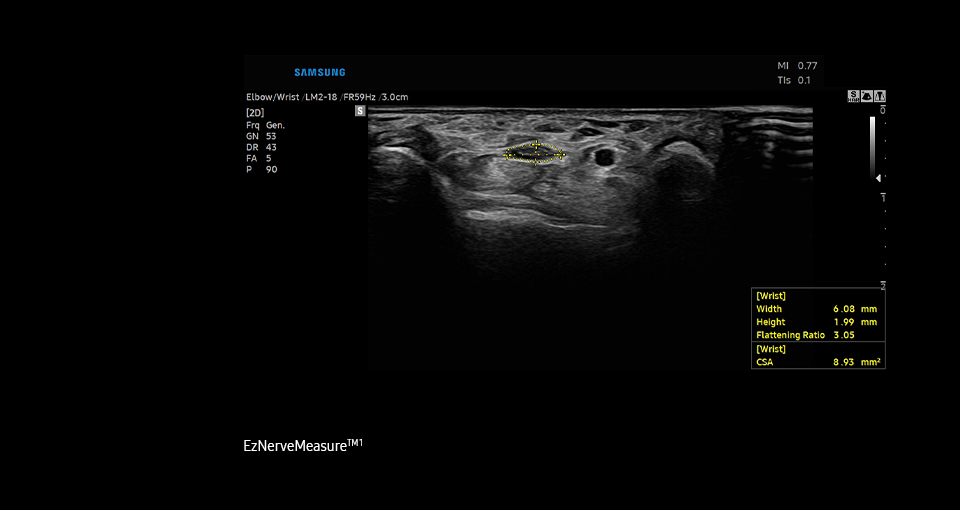
EzNerveMeasure™ ¹ is a feature that provides measurement results
of the long axis, short axis, flattening ratio, and Cross-Sectional
Area of the detected nerve area.
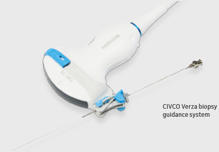
RS85 Prestige provides a broad range of precise fusion, guidance,and dedicated tools to support healthcare professionals strengthen their confidence in operating interventional procedures.
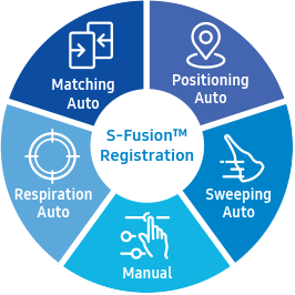
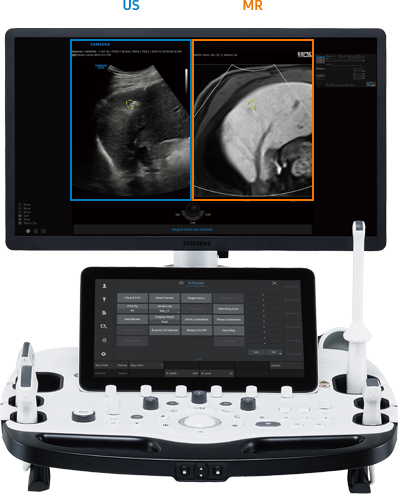
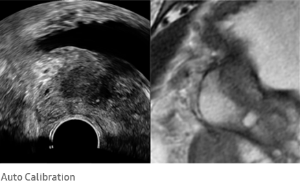
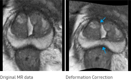
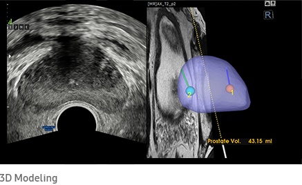
S-Fusion™ ¹ enables simultaneous localization of a lesion using real-time ultrasound in conjunction with other volumetric imaging modalities. Samsung's Auto Registration helps quickly and precisely fuse the images, increasing efficiency and reducing procedure time.
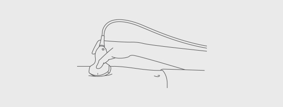
S-Fusion™ ¹ enables precise targeting during interventional and other advanced clinical procedures.

--- --- --- --- --- --- --- --- --- --- --- --- --- --- --- --- --- ---
WHY TO SUBSCRIBE?

Get exclusive deals and offers

Valued content about improving your healthcare business

Newest features & updates
--- --- --- --- --- --- --- --- --- --- --- --- --- --- --- --- --- ---
Related Searches:
#ultrasound iraq #RS85 Prestige #RS85 probes #Samsung #ultrasound Iraq #samsung ultrasound iraq #ultrasound machine #ultrasound scan #ultrasound #Samsung ultrasound distributor
#التراساوند #التراساوند العراق #اجهزة التراساوند #جهاز سامسونك التراساوند #وكيل سامسونك التراساوند في العراق